Congress Speakers
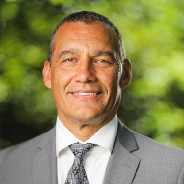
Dr Craig Challen SC OAM

Congress Opening Address
Read bio…
Dr Craig Challen
Dr Craig Challen SC OAM is an Australian cave diver, notable for his efforts in saving a soccer team of twelve boys and their coach in the 2018 Thai Cave Rescue. His unwavering and selfless bravery saw him be awarded with the Star of Courage for the successful rescue of the trapped soccer team.

In 2019, alongside Dr Richard Harris SC OAM, Dr Craig Challen was named as the first dual Australians of the Year for their heroic efforts as part of an international rescue mission to save 12 boys from flooded caves in Thailand.
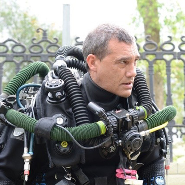
As one of Australia’s leading technical divers, Craig is a member of the Wet Mules, a diving group that takes on some of the world’s deepest underwater caves. After commencing cave diving in the 1990s, he was an early adopter of closed-circuit mixed gas rebreathers.
Craig’s notable explorations include the extension of Cocklebiddy Cave on the Nullarbor Plain of Australia in 2008 and the Pearse Resurgence in New Zealand over the last 10 years. He has additionally explored caves throughout Australia, New Zealand, China, Thailand, Vanuatu and the Cook Islands.
Craig also has an avid interest in shipwreck diving and has explored sites over the last 15 years in the South China Sea, Solomon Islands, Australia, New Zealand, Thailand, Malaysia and Indonesia.
In 2018, Dr Challen, alongside his diving partner Dr Richard ‘Harry’ Harris, were invited to assist in the rescue operation of a Thai soccer team trapped Thailand’s deep Tham Luang Cave, in a story that gripped the world. The pair worked 14-16 hour days making trips into the cave on a daily basis, working cooperatively to ensure the safe evacuation from the cave for everyone. Craig’s expertise and experience in diving saw him help to de-kit the boys of their diving equipment after escaping the first flooded section of the evacuation route, carry them to the next section and prepare them for the next dive.
His unwavering and selfless bravery following the successful rescue of the trapped soccer team saw him be awarded the Star of Courage as well as the Order of Australia for “service to the international community”.
Dr Challen is also a contestant on the current series of ‘SAS Australia’.
Read abstract…
Against all Odds-the story of the 2018 Tham Luang cave rescue
Adventurer, cave diver, veterinary surgeon, businessman and pilot Craig Challen shares his experience as a participant in the 2018 Tham Luang cave rescue, providing an inside account of the extraordinary events that unfolded, focusing on the theme of leadership in a challenging environment.
Craig delves into the crucial role of leadership during the Thai cave rescue, highlighting the vital lessons that can be applied to a variety of contexts, including the medical field. Drawing from his experiences, he emphasises the significance of effective communication, fostering trust within teams, and making tough decisions under extreme pressure. Craig underscores the importance of adaptability, resilience, and maintaining a calm and composed demeanour in demanding circumstances.
The challenges encountered during the cave rescue will be connected to those faced by medical professionals by exploring the critical decision-making processes involved in both scenarios, emphasising the need for thorough evaluation, quick thinking, and the ability to mitigate risks effectively. Craig shares valuable insights on maintaining focus, managing stress, and making calculated choices when lives hang in the balance.
The story of the Tham Luang rescue highlights the universal nature of leadership skills and risk assessment strategies, contributing to a deeper understanding by attendees of the complexities inherent in their daily work and inspiring them to apply the lessons to their own professional challenges. The captivating narrative and practical takeaways make this talk a powerful source of inspiration and guidance for professionals seeking to enhance their leadership capabilities and risk assessment skills, particularly surgeons who face high-stakes situations and engage in risk management during complex procedures.
Dr Jennifer Arnold

Council Lecture
Read bio…
Dr Jennifer Arnold
Dr Jennifer Arnold is a Sydney based medical retinal specialist and researcher.
Trained at the Sydney Eye Hospital followed by a medical retinal fellowship in Paris under Professors Gabriel Coscas and Gisele Soubrane, she worked as a consultant in Aberdeen Scotland for 5 years before returning to Sydney to take up practice at Marsden Eye Specialists.
Working many years with John and Shirley Sarks set the foundation for an ongoing focus in age-related macular degeneration which remains one of her main interests along with retino-vascular diseases.
As well as pursuing full time clinical practice, Jennifer maintains a strong involvement in clinical research resulting in over 80 publications: she runs a dynamic clinical research department and has been principal investigator in over 70 international clinical trials of new treatments for a range of retinal conditions. She is additionally active in the analysis of real world outcomes for the management of retinal diseases as a researcher and committee member of the Fight Retinal Blindness FRB! Programme.
She is widely consulted for local and international advisory boards, research planning and supervising committees, grant application and assessment committees and has recently joined as Medical Advisor to the Vienna-based RetinSight which translates AI programmes into retinal clinical practice. Jennifer is the immediate past chair of the Australian New Zealand Society of Retinal Specialists and is a member of the Macula Society.
Read abstract…
Way points on the journey of a clinical researcher: Where were we then, where we are now, where we are going.
In this Council lecture I will illustrate how clinical research can be combined within an everyday ophthalmic practice to round out a rewarding professional life, describing research of varying methodologies in which I have been involved at all stages of my professional career. I will discuss several examples that exemplify the gradual evolution of knowledge that is incorporated into, and modifies our patient management, as well as pointing to developments and improvements in the future. These examples include: reticular pseudodrusen, macular neovascularisation multimodal imaging and classification, photodynamic therapy, refinements in management of neovascular AMD from clinical trials to clinical practice through real world evidence.
Dr Anthony Bennett Hall

Fred Hollows Lecture
Read bio…
Dr Anthony Bennett Hall
Anthony Bennett Hall was born in Lesotho in Southern Africa. He worked in Zimbabwe and Lesotho as a general medical doctor for a number of years. Anthony spent 12 years in UK, training in general ophthalmology and retinal surgery. He joined CBM in 2000 as head of the Department of Ophthalmology at Kilimanjaro Christian Medical Centre in Tanzania. Based in Tanzania for over a decade, Anthony ran a post graduate programme in ophthalmology and a fellowship programme for Vitreo-retinal surgeons. Anthony is on the board of directors of the Fred Hollows Foundation. He is on the board of Kilimanjaro Centre of Community Ophthalmology (KCCO) Tanzania. He was previously on the Vision 2020 Australia board and served as chairman of RANZCO’s international development committee. he represented RANZCO on the Queen Elizabeth Diamond Jubilee Trust fellowship committee. Anthony is currently a Vitreo-Retinal Surgeon in Newcastle.
Read abstract…
Preventing Blindness from Diabetes in Low and Middle Income Countries- The Diabetic Retinopathy Network(DR-NET)
The number of people with Diabetes Mellitus(DM) in sub-Saharan Africa is projected to increase to about 47 million by 2045. The magnitude of Diabetic Retinopathy (DR) will increase proportionally.
The DR-NET was established in 2014 as part of the VISION 2020 LINKS programme at the International Centre for Eye Health , London School of Hygiene and Tropical Medicine. This eye health partnership programme partners eye units in low and middle income countries (LMICs), with eye units in the UK to improve the quality and quantity of eyecare training and service delivery. There are now 28 DR centres in 20 LMIC including a DR-NET LINK between RANZCO and Pacific Island programmes.
The DR-NET key activities included, situation analysis of partners, workshops bringing together whole eye teams including the MoH representative, development of 2 year action plans, A DR-NET toolkit to plan and implement services, whole team training visits between LINKS partners to build capacity and the development of national DR guidelines in collaboration with MOH.
Kilimanjaro Christian Medical Centre in Tanzania was one of the first LINKS programmes. We established a screening programme for DR using an intervention mapping approach. Key aspects of this programme included a needs assessment of people living with diabetes (PLWD) and health care workers, a trial of DR screening methods, comic strips as motivational strategy to increase uptake of DR screening, healthcare worker education, an electronic database of PLWD, implementation of mobile DR screening and programme evaluation.
The Fred Hollows Foundation is leading research in innovative approaches to DR screening.
The LINKS programme has proved to be a mutually beneficial way of building capacity to treat and prevent unnecessary loss of sight and blindness in LMICs. There remain enormous challenges ahead to eliminate avoidable visual impairment particularly from DR.
Prof Kathryn Burdon

Ida Mann Memorial Lecture
Read bio…
Professor Kathryn P Burdon
Kathryn Burdon began her journey in ophthalmic genetics with a PhD from University of Tasmania in 2004 on the genetics of paediatric cataract where she discovered the genetic cause of Nance-Horan Syndrome. She spent two years as a post-doctoral fellow at Wake Forest University Baptist Medical Center in North Carolina, USA, working on complex disease genetics, primarily the cardiovascular and renal complications of diabetes mellitus. On her return to Australia in 2005 she joined the Department of Ophthalmology at Flinders University in Adelaide where she established a research group focused on the identification of genes for blinding diseases, including glaucoma, keratoconus and diabetic retinopathy, as well as continuing her work in childhood cataract.
In 2014 she returned to the University of Tasmania to join the genetics theme at the Menzies Institute for Medical Research where she is now the Theme Leader. She runs an internationally recognised ophthalmic genetics research program focused on identifying the genetic risk factors for blinding eye disease and characterising the causative genes and variants in animal and cellular models. She has skills in molecular biology, human genetics, genetic statistics and bioinformatics and collaborates broadly with ophthalmologists and geneticists around the world.
Read abstract…
Why is my child blind? How genomics provides the answers patients and parents seek.
There are over 900 recognised rare eye diseases, and most of them are caused by small changes to a big genome. The overarching goal of my research program is to generate the knowledge that allows us to provide accurate genetic diagnosis for rare eye disease through identifying and characterising the causative genes and genetic variants. Recent leaps forward in technological capability for interrogating the human genome have driven an explosion in new knowledge about the genes and variants that cause hundreds of rare eye diseases. In addition to discovering new genes, we have found that the genetic and molecular underpinnings of disease do not always reflect our clinical classifications, and that the genetic heterogeneity of many rare diseases is immense. Our ability to interrogate entire genomes has revolutionised both gene discovery and diagnostics but has also highlighted the challenges of interpreting genetic data in individuals, kickstarting international efforts to characterise and categorise genetic variants. This presentation will explore the benefits of genetic diagnosis for patients and families, illustrating modern approaches to gene discovery for rare eye disease and highlight important contributions Australia is making to global efforts to understand the clinical impacts of genetic variants. Advances in genomic technologies and understanding of disease-causing genes continue to increase the utility of genomic testing in the diagnosis and management of rare eye diseases.
A/Prof Fred Chen

Retina Update Lecture
Read bio…
A/Prof Fred Chen
Dr Fred Chen is a vitreoretinal surgeon and clinician-researcher based in Perth. In 2010, after completing both a PhD at the University College London in retinal cell transplantation and medical and surgical retina Fellowships at Moorfields Eye Hospital, Dr Chen returned to Perth to establish the Ocular Tissue Engineering Laboratory at the Lions Eye Institute to further the investigation of stem cell therapy and inherited retinal diseases.
Dr Chen is also an active principal investigator in ophthalmic clinical trials of novel retinal therapies and systemic drug toxicity, his clinical interest being the natural history of genetic eye diseases and clinical trial endpoints. His laboratory examines variants of uncertain significance, using patient-derived cell models, and also develops gene replacement and patient-customised antisense therapies that alter gene splicing to treat inherited retinal diseases.
Dr Chen has been awarded numerous fellowships and research grants from the Australian National Health and Medical Research Council. He is a Fellow of the Royal Australian and New Zealand College of Ophthalmologists and has previously served on the Board of the Ophthalmic Research Institute of Australia. He is a Principal Research Fellow at the University of Western Australia and a Clinical Associate Professor at the University of Melbourne.
Read abstract…
Deep phenotyping, pitfalls in pivotal trials and precision genetics.
Multimodal retinal imaging is now the standard of care for retinal diagnostics and management. Cases will be presented to illustrate the essential role multimodal imaging plays in the work-up of complex clinical scenarios. The use of artificial intelligence, through incorporating large data sets from research and clinical care settings for deep learning, is now gaining momentum. Although issues with implementation remain, potential solutions are on the horizon.
New agents for the treatment of neovascular AMD and geographic atrophy have now been approved by the major regulatory agencies for routine clinical care. Do we know enough, however, about the efficacy of these therapies to embrace them? Do these new treatments, lacking long term safety data, offer significant gains over our current armamentarium? For geographic atrophy, is there sufficient trial data to support the widespread use of complement inhibitors? New methods of drug delivery for AMD, DMO and macular telangiectasia type 2 will be covered.
Our surgical and medical retinal subspecialities are now being transformed by a tidal wave of rapid advances in precision medicine. Molecular diagnostics is gaining traction for accurate diagnosis and prognosis within the fields of ocular oncology and inherited retinal diseases. This technology, however, is not infallible. Understanding the pitfalls is important given the development of personalised gene therapy. This emergence of molecular medicine will be illustrated by the utility of circulating tumour DNA in the management of choroidal melanoma and the Australian led development of antisense therapy for Retinitis Pigmentosa type 11.
Dr Shigeru Kinoshita

Sir Norman Gregg Lecture
Read bio…
Dr Shigeru Kinoshita
Dr Shigeru Kinoshita, a clinician scientist, graduated from Osaka University Medical School in 1974, and has served as the Professor and Chair of Ophthalmology at Kyoto Prefectural University of Medicine since 1992. Because of his stepping down from the Chair of Ophthalmology in March 2015, He was elected the Professor and Chair of Frontier Medical Science and Technology for Ophthalmology at Kyoto Prefectural University of Medicine in April 2015. And, he has been continuously working as a distinguished clinician scientist.
In the early 1980s at Harvard Medical School, he, in collaboration with Dr. Richard A. Thoft, established the concept of centripetal movement of corneal epithelium, and his groundbreaking work has shed new light on the importance of limbal epithelium. His series of findings has had an enormous impact on this subject and has afforded much insight, ultimately contributing to the development of the corneal stem cell theory set forth by Tuen-Tien Sun. Based on these concepts, Dr. Kinoshita developed a new surgical procedure for in vivo corneal epithelial transplantation that has led to epithelial stem cell transplantation for ocular surface rehabilitation. Over the past 40 years, his primary interest has been focused on the translational research of new therapeutic modalities for severe corneal diseases. Following this path, his group has established the rational design and technologies of cultivated mucosal epithelial stem cell transplantation for severe ocular surface disorders such as Stevens-Johnson syndrome and chemical injury, and cultivated human corneal endothelial cell (CHCEC) injection therapy for corneal endothelial dysfunction. His group also proved the clinical efficacy of Rho-associated protein kinase (ROCK)-inhibitor topical application for partial corneal endothelial dysfunction.
Kinoshita is a recipient of the 1999 Alcon Research Institute Award, the 2008 Castroviejo Medal Lecturer of the Cornea Society, the 2009 ARVO Gold Fellow, the 2010 Claes H. Dohlman Conference Address of the TFOS, the 2010 Meibom Lecturer in Germany, the Doyne Memorial Lecturer of the 2011 Oxford Ophthalmological Congress in United Kingdom, the 2011 Elsemay Bjorn Lecture in Finland, Schepens Eye Research Institute Alumnus Awardee 2011, the Peter Herberg Lecture at IMCLC2012, the Richard Lindstrom Lecture, CLAO, ASCRS 2014, Charles D. Kelman Innovator Award, ASCRS 2015, the Friedenwal Award Lecturer at the ARVO 2016, The Coster Lecture at the Australian and New Zealand Cornea Society 2017, and David Easty Lecture at the Bowman’s Club 2017. He served as an ARVO Program Committee Member in the Cornea Section between 1996 and 1999, the ARVO Trustee of the Cornea Section between 2006 and 2011, and the ARVO Vice President in 2010-2011.
Read abstract…
Toward Corneal Regenerative Medicine
Devastating ocular surface and cornea-related disorders, such as Stevens-Johnson syndrome, chemical injury, Fuchs endothelial corneal dystrophy, and severe corneal endothelial failures, are very difficult to treat properly. Several types of transplantable cultivated mucosal epithelial sheets have been developed thanks to state-of-the-art corneal regenerative medicine and the latest advancements in ocular surface biology. The first is the allogeneic/autologous corneal epithelial stem-cell sheet, the second is the autologous oral mucosal epithelial sheet, and the third is the iPS-cell derived corneal epithelial sheet. Some of them have been officially approved by the EMA and the PMDA for clinical use.
A similar corneal regenerative medicine can be applied to treat corneal endothelial dysfunction. For example, a surgical modality using novel cultured human corneal endothelial cells (CHCEC) which is the injection of CHCEC with ROCK inhibitor into the anterior chamber has now shown promise in clinical efficacy. Another aspect of our cutting-edge translational research is focused on developing a novel medical treatment for the early-phase corneal endothelial disease. To that end, Rho-associated protein kinase (ROCK)-inhibitor eye drops have proved effective in treating partial endothelial dysfunction.
It is our great hope that cornea-related translational research, such as that described above, will receive official governmental approval based on the accumulated data on the safety and efficacy aspects of the procedures, thus ultimately resulting in the worldwide prevention of blindness.
Vincenzo Maurino MD

Cataract Update Lecture
Read bio…
Vincenzo Maurino MD, BQOphth, CertLAS RCOphth
Vincenzo Maurino is an Italian-British ophthalmologist who is Consultant Ophthalmologist and Director of the Cataract Service at Moorfields Eye Hospital in London. His special areas of interests are cataract surgery and corneal surgery as well as refractive surgery.
He graduated with magna cum laude in Italy and after a four-year ophthalmology residency in Italy, he won a scholarship to move to London to further his surgical ophthalmic training. At Moorfields Eye Hospital he completed fellowships in paediatric ophthalmology, glaucoma, cataract, refractive surgery, and corneal transplant surgery.
Vincenzo Maurino research interests lie in the fields of cataract and corneal surgery, and he has published in several peer reviewed international journals. He has been invited to lectures nationally and internationally. He served on the scientific committee of the Italian Society of Ophthalmology (SOI) for longer than a decade and on the board of the SICCSO (The International Society of Corneal, Stem Cells and Ocular Surface). He was awarded the SOI Medal Lecture in 2016. He received the title of Officer of the Order of The Star of Italy by Mr Sergio Mattarella, the President of Italy, in 2015. He is passionate about surgical training and has trained hundreds of eye surgeons from all over the world in cataract and corneal surgery and he regularly holds training surgical courses nationally and abroad. He has continued working in Italy as Visiting Professor of corneal surgery at the Tor Vergata University in Rome.
Consultant Ophthalmic Surgeon, Corneal and Refractive Surgery Department
Director Cataract Surgical Service
Moorfields Eye Hospital
London UK
Read abstract…
Challenging cataract surgery and new trends in cataract surgery.
Cataract surgery is never to be underestimated and remains a complex eye procedure that requires experience, practice, and surgical situational awareness to be successfully completed. I will discuss what is involved in my daily cataract practice and how I manage it and latest changes to my cataract surgery.
- Complex cataract cases especially brunescent cataract and cataract with zonulopathy need extra and specific counselling. Detailed planning is needed to approach these cases with confidence and safety and achieve success. I will discuss and provide video examples of the approach to different challenging brunescent cataract and cataract with zonulopathy and their management.
- High volume cataract surgery and the advent of ISBCS (Immediate Sequential Bilateral Cataract Surgery) that goes alongside high-volume surgery. ISBCS is here to stay and to become the norm due to its advantages and minimal/similar risks to sequential surgery.
- Cataract surgery is now a viable way to achieve spectacle independence in our older patients with the latest EDOF (Extended Depth of Focus) IOL causing minimal visual side effects compared with previous generation multifocal diffractive IOL.
- IOL implants are not risk free and some IOL have recently been plagued by problems with severe IOL mineralisation causing complete IOL opacification. I will show how to tackle these cases and the techniques used to minimise zonular damage and to enable “in the bag” or sulcus IOL exchange and enhance patients safety and outcome.
Dr Neil Miller

Neuro-ophthalmology Update Lecture
Read bio…
Neil R. Miller, MD, FACS
Dr Neil Miller is the Frank B. Walsh Professor of Neuro-Ophthalmology and Professor of Ophthalmology, Neurology, and Neurosurgery at the Johns Hopkins University School of Medicine. He has authored over 570 articles, 93 chapters, and 13 books, including the 4th edition of “Walsh and Hoyt’s Clinical Neuro-Ophthalmology” and has co-edited the 5th and 6th editions of this textbook as well as four editions of an abbreviated version of the textbook: “Walsh and Hoyt’s Clinical Neuro-Ophthalmology: The Essentials”, the most recent of which was published in 2020.
Dr. Miller also has co-authored two editions of “The Neuro-Ophthalmology Survival Guide”, a textbook designed for both physicians and students. Dr. Miller has spoken at numerous local, national, and international meetings and has given 62 named lectures around the world. In addition, he has been involved with many clinical trials in the field of neuro-ophthalmology. Many of Dr. Miller’s previous Fellows and residents hold faculty positions at major institutions throughout the United States and around the world.
Read abstract…
Neuro-Ophthalmology Updates: Information That Will Change Your Practice Tomorrow!
Recently, there have been a number of important advances in the diagnosis and management of several neuro-ophthalmic disorders. These advances have major implications for practitioners and should change your practice if you have not changed it already! In this talk, I will discuss several of what, in my opinion, are the most important. These include advances that have changed the approach to and management of patients with acute optic neuritis. Specifically, it is no longer appropriate to offer all patients with acute optic neuritis the “option” of treatment with systemic corticosteroids. Instead, all patients except for those suspected of having an infectious etiology (eg, tuberculosis) should be treated immediately with high-dose steroids. In addition, patients with recurrent steroid-sensitive but steroid-dependent optic neuritis require an assay for antibodies to myelin oligodendrocyte glycoprotein. With respect to idiopathic intracranial hypertension (aka primary pseudotumor cerebri), we now know that patients with this condition can tolerate up to 4 grams of acetazolamide per day and that permanent weight loss—a major objective in the successful management of such patients—can be better achieved with bariatric surgery than through weight loss clinics. It is now clear that “visual snow” is an organic disorder of perception and should be treated as such rather than as a psychological problem. Finally, molecular genetics is playing an increasingly important role in the management of children with optic pathway gliomas, so much so that targeted therapy is now both available and beneficial for such individuals.
Prof Dan Reinstein

Refractive Update Lecture
Read bio…
Professor Dan Z. Reinstein
He has delivered over 1,000 lectures at professional meetings on 5 continents and published over 200 articles in peer-reviewed medical journals. He is a leader in the field of Therapeutic Refractive Surgery, and founded this section for the Journal of Refractive Surgery where he continues as Section Editor. He has authored a definitive textbook on SMILE, contributed to 44 book chapters and published proceedings, and is extensively published in the ophthalmic press.
His work and patents related to VHF digital ultrasound administered by the Center for Technology Licensing at Cornell University (Ithaca, NY) led to the commercialisation of VHF digital ultrasound robotic scanning with the Insight 100 from ArcScan Inc., which together with his sizing formula is now the most accurate method of sizing the ICL. He developed PRESBYOND Laser Blended Vision, now part of the Carl Zeiss Meditec platform. He has been the Lead Refractive Surgery consultant for Carl Zeiss Meditec since 2001, has a proprietary interest in the Insight 100 technology, and consults for CSO Italia (MS39 OCT).
Read abstract…
What innovations is a practice offering to be at the forefront of refractive surgery?
Three major innovations have reached maturity from the forefront of refractive surgery to be generalised amongst refractive surgeons: SMILE, PRESBYOND and ICL sizing.
– SMILE, with its unique biomechanical profile, enables larger optical zones to be programmed resulting in less spherical aberration induction and hence better night vision, while better preserving corneal nerves giving less dry eye symptoms and more rapid return to normal, including contact sports. The VISUMAX 800 introduced in 2021, provides technical improvements including 3x faster lenticule cutting time (<10s) and software control for centration and cyclotorsion optimizing both the efficacy and safety of treatment.
– PRESBYOND is a corneal treatment option for presbyopia combining a micro-anisometropia with extended depth of field (EDoF) produced by modulation of spherical aberration. PRESBYOND enables the correction of plano presbyopia and astigmatic ametropias between -8.00D to +5.00D. This modality obviates the need for the higher risk and lower accuracy of clear lens extraction. With over 97% of the population as suitable candidates, the bilateral LASIK procedure enabling a prompt return to work and daily activities with less side effects, faster adaptation time and no loss of contrast. LASIK accuracy offers improved SEQ and cylinder accuracy as well as future adjustability, reversibility and an in-built EDoF that enables high quality optic, low PCO rate monofocal IOLs to be used for future cataract surgery if needed, without resorting to contrast-lowering multifocal or diffractive IOLs.
– ICL has significantly expanded the treatment range outside of that amenable to corneal refractive surgery. However, the challenge of ICL sizing problems requiring exchange or more serious complications is probably responsible for generally low adoption rates (excluding where marketing and social engineering leads to ICL implantation despite being excellent candidates for corneal procedures). Very high-frequency digital ultrasound (VHFDU) and the Reinstein ICL formula has produced significantly better sizing with vault prediction improved by a factor of 3.4 compared to white-to-white and by a factor of 2.2 compared to anterior segment OCT based sizing. By Reinstein formula 61% of eyes achieve a vault within ±100μm and 96% within ±300μm of predicted. Thus VHFDU sizing provides greatly improved early and long-term safety for ICL technology making it potentially an alternative to corneal refractive surgery for the general ophthalmologist that has not invested in laser technology and specialist training to perform corneal refractive surgery.
Prof Tina Wong

Glaucoma
Read bio…
Professor Tina Wong
Head of the Glaucoma Service and Senior Consultant at the Singapore National Eye Centre, Director of Clinical Translational Research and Head of the Ocular Therapeutics and Drug Delivery Group at the Singapore Eye Research Institute and Professor at DUKE-NUS Graduate Medical School. Professor Wong is the founding President of the Glaucoma Association Singapore. She is the Executive Director of the National Health Innovation Centre supporting healthcare innovation and enterprise in Singapore.
Professor Wong’s research focuses on the development of new therapeutics to improve surgical and clinical outcomes. She is an internationally renowned glaucoma specialist, with an interest in ocular wound healing and post glaucoma surgery management. She leads an interdisciplinary research programme on translational and clinical research on ocular wound healing and drug delivery.
Professor Wong’s research has improved the understanding of the burden and risk factors that affect the surgical outcomes of glaucoma surgery. She has developed 2 sustained drug delivery therapies, reaching clinical trials for the medical treatment of glaucoma and cataract surgery. She holds 10 patents to her name and is a co-founder of 2 spin off biotech companies and have attracted several millions of dollars of private investment. Professor Wong is a strong advocate for translating innovative research to healthcare solutions that will directly improve quality and access to care.
Professor Wong is a scientific thought leader in the field of wound healing in glaucoma surgery and ocular drug delivery, having published widely in these areas, with Google scholar H-index of 42. She has nationally funded research grants amounting to SG$25M. She has received national and international awards for her contribution to the field of glaucoma surgical wound healing and ocular drug delivery. Of notable mention, Professor Wong was awarded the President’s Science and Technology Award in Singapore in 2014. This award is the country’s highest honour bestowed on exceptional research scientists and engineers in Singapore for their excellent achievements in science and technology. These national awards are given annually to recognise and celebrate outstanding and invaluable contributions by individuals or teams to the research and development landscape in Singapore. This research was recognized as one of the top 50 significant developments in Singapore in past 50 years in the same year.
Read abstract…
Making Blebs Beautiful Again: Insights into Collagen Remodelling and the Future Direction of Anti-Fibrotic Strategies.
Surgical options have long been historically reserved for patients with either advanced stages of the disease and/or ineffective IOP control with medications or laser associated with signs of progressive loss of vision. Trabeculectomy surgery is a highly effective operation in lowering the IOP but can have risks of failure mainly from the wound healing response and scarring as well as a lifetime risk of blindness as a result of MMC prescribed as the Gold standard of care since the late 1980s. Whilst over the past 4 decades we have witnessed tremendous improvements in surgical techniques, as well as more recently, the emergence of minimally invasive devices further adding to our surgical armamentarium, the anti-fibrotic management in bleb forming glaucoma surgeries have largely remained stagnant and unchanged. With the increasing life span in the world population and greater accessibility to surgery at earlier stages of disease, we are witnessing a significant increase in the number of patients undergoing surgery. For most countries, bleb forming surgeries remain an important surgical option for achieving target IOPs of low teens to single digits.Unpredictable scarring is a serious complication that hampers success in achieving the goal of long-term IOP control. Finding an alternative to MMC remains elusive and drives continued efforts interrogating the biological processes fuelling this obstacle. In this lecture, new emerging concepts and ongoing research directions will be discussed that are striving to deliver safer anti-scarring strategies.